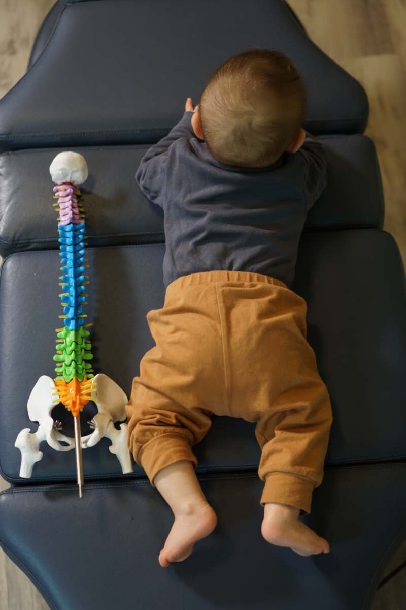
Corrective exercises enhance joint mobility by addressing neuromuscular dysfunction, restoring optimal movement patterns, and increasing tissue extensibility through targeted motor control training. Unlike passive stretching, these evidence-based protocols reprogram how your nervous system coordinates movement, eliminating compensatory patterns that restrict range of motion. When properly prescribed and progressed, corrective exercises create lasting mobility improvements by combining strength, stability, and controlled movement transforming how your joints function during everyday activities and athletic performance.
The Neuromuscular Mechanism Behind Corrective Exercise-Driven Mobility Gains
Understanding how do corrective exercises improve joint mobility requires looking beyond simple stretching mechanics. The real magic happens at the nervous system level. Your brain constantly monitors joint position and muscle tension through proprioceptive feedback loops. When dysfunction occurs, these protective mechanisms actually work against you, creating what clinicians call neuromuscular inhibition.
This inhibition manifests as muscles that won’t fully activate despite your best efforts. You might stretch a tight hip flexor for months with minimal results because the opposing glute muscles aren’t firing properly. The hip flexor remains tight as a compensatory stabilization strategy. Traditional stretching addresses symptoms but misses the root cause entirely.
Motor control theory explains that movement patterns become hardwired through repeated practice. Poor posture, previous injuries, or sedentary lifestyles create dysfunctional patterns that your nervous system perceives as “normal.” Corrective exercises work by interrupting these maladaptive loops and establishing new neuromuscular pathways. Research from 2026 demonstrates that targeted motor control training can reestablish optimal recruitment sequences within weeks when practitioners apply proper progression protocols.

The nervous system’s plasticity allows for remarkable adaptation when given appropriate stimuli. Progressive challenges that demand precision and control force the brain to upgrade its movement software. This neuroplastic adaptation explains why improved mobility through chiropractic care often requires active patient participation rather than passive treatment alone.
| Outcome Measure | Passive Stretching | Corrective Exercise |
|---|---|---|
| Duration of Effect | Minutes to hours | Weeks to months |
| Mechanism | Temporary tissue elongation | Neuromuscular reprogramming |
| Strength Integration | Minimal | Full activation required |
| Compensatory Pattern Addressed | No | Yes |
| Functional Carryover | Limited | High |
Quick Answer: Three Primary Ways Corrective Exercises Restore Joint Function
For those seeking immediate clarity on the mechanisms at play, three distinct pathways explain how targeted movement interventions translate into measurable mobility gains:
- Motor Pattern Correction: Exercises retrain the nervous system’s coordination sequences, replacing dysfunctional movement habits with efficient patterns that allow full joint range of motion without compensatory restrictions.
- Tissue Remodeling: Controlled loading through progressive exercises stimulates collagen synthesis and realignment, improving tissue extensibility while building structural resilience against future restriction.
- Proprioceptive Enhancement: Targeted movements heighten joint position awareness, reducing protective guarding responses that unnecessarily limit mobility and enabling confident movement through full ranges.

Each mechanism builds upon the others. Better motor control enables proper loading, which drives tissue adaptation. Enhanced proprioception reinforces improved patterns, creating an upward spiral of functional mobility. Patients engaging with rehabilitation exercises often notice improvements compound over time as these systems reinforce each other.
Movement Assessment Protocols That Identify Mobility-Limiting Dysfunction
Effective intervention begins with accurate diagnosis. Not all joint restrictions feel the same, and distinguishing between structural limitations and functional compensations determines treatment success. Modern movement screening has evolved significantly, with validated protocols providing objective baselines for tracking progress.
The Functional Movement Screen (FMS) and its clinical counterpart, the Selective Functional Movement Assessment (SFMA), represent gold-standard approaches for identifying dysfunctional patterns. These systematic evaluations break down complex movements into component parts, revealing where the kinetic chain breaks down. A patient might demonstrate adequate isolated hip flexion but fail to integrate that mobility during a compound movement like a deep squat.
Compensation patterns mask true restrictions cleverly. Someone lacking thoracic spine extension might hyperextend their lumbar spine instead, creating lower back pain while the actual stiff segment goes unaddressed. Without proper screening, treatment targets the painful area rather than the responsible dysfunction.
Clinical studies show that movement screening identifies dysfunction patterns that traditional orthopedic examinations miss. The value lies in assessing integrated movement rather than isolated joint function. After all, nobody moves one joint at a time outside the treatment room.
Research indicates that movement screening protocols identify underlying dysfunction in approximately 70% of patients presenting with mobility complaints who had previously received treatment addressing symptoms rather than causes.
Distinguishing tightness from weakness requires hands-on assessment combined with movement observation. A muscle might feel tight because it’s overworking to compensate for inhibited synergists. assisted stretching for mobility can provide temporary relief, but lasting change requires addressing why that muscle works overtime in the first place.
How to Execute Progressive Corrective Exercise Programs for Sustained Mobility
Transitioning from assessment to intervention demands a systematic approach. Random exercises, even well-chosen ones, rarely produce lasting change without proper progression. The following framework guides practitioners and patients through effective implementation:
- Baseline Assessment: Establish objective mobility measurements using standardized tests. Document both passive range and active control throughout that range, noting any pain provocation or compensatory patterns during assessment.
- Priority Identification: Determine which dysfunctions create the greatest functional limitation. Address proximal stability before distal mobility, and resolve distal restrictions before challenging proximal control.
- Foundational Activation: Begin with isolated activation exercises for inhibited muscles. Use supported positions that minimize compensatory recruitment before progressing to integrated movements.
- Pattern Integration: Once individual components activate properly, combine them into functional patterns. Start with slow, controlled movements before adding speed or load.
- Progressive Loading: Gradually increase challenge through range of motion demands, eccentric control, variable surfaces, or external resistance. Document progression criteria and reassess regularly to ensure adaptation continues.
Tissue adaptation timelines vary based on individual factors, but generally follow predictable patterns. Neuromuscular changes emerge within the first few weeks as motor learning accelerates. Structural tissue changes require six to twelve weeks of consistent loading. Patients pursuing hip range of motion improvements should expect this progression timeline when committing to corrective protocols.
Joint-Specific Corrective Strategies: Hip, Shoulder, and Thoracic Mobility Protocols
Different joints present unique challenges requiring region-specific approaches. The hip, shoulder, and thoracic spine commonly restrict mobility, yet each demands distinct corrective strategies based on anatomical and functional characteristics.
Hip mobility limitations often stem from prolonged sitting and anterior-dominant activities. The psoas and iliacus adapt to shortened positions, while gluteal muscles become inhibited from constant lengthening. Effective hip mobility protocols must address both the anterior hip flexors and posterior chain activation simultaneously. Failure to address both sides of the joint perpetuates compensatory strategies.
Shoulder complex dysfunction requires analyzing the scapular-thoracic relationship alongside glenohumeral movement. Many apparent shoulder restrictions actually reflect poor scapular positioning. The thoracic spine must extend adequately for the scapula to upwardly rotate, allowing overhead mobility. Treating the glenohumeral joint in isolation ignores this critical proximal relationship.
Thoracic mobility deficits create cascading effects throughout the kinetic chain. Limited extension forces cervical and lumbar compensations. Restricted rotation transfers excessive torsional demands to the lumbar spine and shoulders. Addressing thoracic mobility often resolves distal complaints that seemed unrelated.
| Joint Region | Primary Dysfunction | Corrective Exercise Category | Progression Level |
|---|---|---|---|
| Hip | Anterior capsule restriction | Active hip flexor distraction with glute activation | Foundational |
| Hip | Weak posterior chain | Single-leg hip hinge variations | Intermediate |
| Shoulder | Scapular dyskinesis | Quadruped scapular clocks | Foundational |
| Shoulder | Glenohumeral internal rotation deficit | Side-lying internal rotation with distraction | Intermediate |
| Thoracic | Extension limitation | Prone extension over foam roller | Foundational |
| Thoracic | Rotation restriction | Quadruped thread-the-needle with reach | Intermediate |
Data from 2026 clinical outcomes indicates that region-specific protocols outperform generalized mobility routines by significant margins. Patients combining targeted exercises with SI joint stabilization exercises demonstrate superior outcomes compared to those focusing on isolated mobility work alone.
Tissue Extensibility vs Neuromuscular Control: Understanding the Mobility Equation
Not all tightness responds to the same intervention. Distinguishing between structural limitations and functional restrictions prevents wasted effort and accelerates results. Understanding this distinction fundamentally changes how practitioners approach mobility work.
Structural limitations involve actual tissue shortening capsular contractures, shortened muscle-tendon units, or accumulated scar tissue. These restrictions persist regardless of positioning, warm-up status, or mental state. Palpation reveals dense, fibrotic tissue quality. Addressing structural restrictions often requires manual intervention alongside corrective loading.
Functional restrictions fluctuate. The same movement might feel restricted in the morning but improve after activity. Stress, fatigue, or pain perception alters available range. These variations indicate neuromuscular guarding rather than true tissue limitation. Functional restrictions respond beautifully to corrective exercises that rebuild motor control confidence.
Manual therapy accelerates results for both categories but for different reasons. For structural restrictions, hands-on work addresses tissue density directly. For functional limitations, skilled touch provides proprioceptive input that helps recalibrate the nervous system’s threat perception. FAKTR soft tissue therapy exemplifies this dual-purpose approach.
Clinical analysis reveals that approximately 60% of patients presenting with mobility restrictions demonstrate primarily functional limitations, meaning their nervous system restricts range as a protective mechanism rather than actual tissue shortening preventing movement.
Testing helps differentiate. Pass the knee to chest test: if passive hip flexion exceeds active hip flexion by more than 15 degrees, neural inhibition likely limits your mobility. Address motor control first. If passive and active ranges match but remain restricted, structural elements require attention.
Evidence-Based Programming Variables That Maximize Joint Mobility Outcomes
Even the perfect exercise fails without proper programming parameters. Research consistently demonstrates that frequency, intensity, and progression variables determine whether corrective protocols succeed or stagnate. The following principles guide effective prescription:
- Frequency Matters More Than Duration: Brief, frequent practice sessions outperform marathon mobility sessions. Nervous system learning requires repetition with adequate recovery between efforts. Daily ten-minute sessions trump weekly hour-long routines.
- Intensity Requires Precision: Progressive challenge must remain within the range the nervous system can control. Pushing into compensation reinforces the patterns you’re trying to eliminate. Moderate intensity that demands attention without overwhelming capacity drives adaptation.
- Volume Follows Tolerance: Start with fewer repetitions performed perfectly. Adding volume before establishing proper patterns merely reinforces dysfunction. Three pristine repetitions outperform thirty sloppy ones.
- Temporal Positioning Affects Integration: Performing corrective exercises before training primes proper patterns for the workout. Post-training sessions address mobility limitations uncovered by activity. Both serve distinct purposes.
- Progression Criteria Prevent Plateaus: Objective benchmarks determine readiness for advancement. Subjective feelings often mislead. Establish measurable criteria for increasing challenge.
- Variability Enhances Transfer: Once established, vary the context of practiced patterns. Different surfaces, speeds, and loads ensure the nervous system generalizes improved mobility beyond the exercise setting.
Common programming errors sabotage results. Many patients progress too quickly, adding challenge before foundational patterns stabilize. Others abandon exercises when immediate results disappoint, unaware that neuromuscular adaptation requires consistent practice. Structural changes demand patience beyond what most expect.
Regression remains underutilized. When form deteriorates, returning to easier variations rebuilds proper patterns. Stubborn adherence to progressed exercises despite compensatory movement perpetuates dysfunction. Smart programming includes clear regression criteria alongside progression benchmarks.
Those focused on posture correction benefits often underestimate the programming complexity required. Postural muscles require different stimulus parameters than prime movers. Endurance emphasis, lower loads, and higher repetition ranges develop postural endurance without triggering protective tension.
Key Takeaways: Corrective Exercise Integration for Long-Term Mobility Health
Lasting mobility requires systematic intervention rather than scattered exercises. The principles outlined throughout this article coalesce into actionable frameworks that patients and practitioners can implement confidently. The following essential points guide effective corrective exercise programming:
- Assessment Precedes Intervention: Understanding whether restrictions stem from structural limitations or functional compensations determines appropriate strategy selection.
- Neuromuscular Reeducation Drives Change: Passive interventions alone rarely produce lasting results because the nervous system must learn to control newly available range of motion.
- Progressive Loading Builds Resilience: Tissue adaptation requires controlled challenge that progressively exceeds current capacity without overwhelming recovery ability.
- Proximal Stability Enables Distal Mobility: Addressing core and hip stability often resolves extremity restrictions that resisted targeted treatment attempts.
- Consistent Practice Outperforms Intensity: Brief, frequent sessions establish motor patterns more effectively than occasional marathon mobility sessions.
Personalized care ensures protocols match individual needs rather than generic templates. The practitioners at The Chiropractorr emphasize comprehensive assessment and individualized progression based on each patient’s unique presentation, goals, and response patterns. For those seeking to understand mobility’s impact on daily function, recognizing this individualized approach allows for realistic expectations and sustainable implementation.
FAQ: Corrective Exercises and Joint Mobility Enhancement
How long does it take to see mobility improvements from corrective exercises?
Yes, but timelines vary based on restriction type. Neuromuscular adaptations emerge within two to four weeks of consistent practice, while structural tissue changes require eight to twelve weeks. Functional restrictions respond faster than structural limitations that developed over years.
Should corrective exercises cause pain or discomfort?
No, pain indicates incorrect execution or inappropriate progression. Proper corrective exercises create awareness and mild challenge without pain. Discomfort exceeding three out of ten on a pain scale suggests compensation or guarding rather than productive intervention.
Can I do corrective exercises on my own or do I need supervision?
Both, but initial guidance prevents wasted effort. Professional prescription ensures proper exercise selection and technique. Patients demonstrating competency can transition to independent practice, with periodic reassessment confirming continued progress and proper form maintenance.
Do I need to continue corrective exercises forever?
No, but maintenance varies by individual situation. Most patients transition from corrective to performance-based training once dysfunction resolves. Those with chronic conditions or demanding activities may benefit from ongoing maintenance sessions performed weekly rather than daily.
What’s the difference between corrective exercises and regular stretching?
Corrective exercises address root causes rather than symptoms. Traditional stretching temporarily increases tissue length without addressing why restriction exists. Corrective exercises retrain neuromuscular patterns, creating lasting change through motor control rather than passive elongation.
Can corrective exercises help with joint pain or just stiffness?
Yes, pain often decreases as movement quality improves. Many joint pain complaints stem from compensatory patterns and abnormal loading. Restoring proper movement mechanics reduces irregular stress on sensitive structures, addressing the mechanical source of discomfort alongside mobility limitations.